elocation-id: e4251
When Agave angustifolia stem tissues are established in vitro, microbial contamination frequently occurs; therefore, the use of antibiotics to solve this problem was evaluated. The purpose of this work was to determine the percentage of stem tissues that had an aseptic establishment and exhibited morphogenetic response in three situations: 1) during in vitro establishment (stage I); 2) in explants in which organogenesis with shoot formation occurred in stage I but were contaminated with bacteria, and these were treated with antibiotics to eliminate contamination; and 3) in aseptic adventitious shoots obtained in stage I and transferred to propagule multiplication medium (stage II). The percentages of aseptic cultures, viable tissues, and organogenic response were evaluated. The results showed that only between 3.68 and 8.73% of stem tissues were aseptic during stage I. However, by applying antibiotics to shoots already formed but contaminated with bacteria at this stage, asepsis was recovered in 52.5% of the cultures. On the other hand, adventitious shoots that were aseptic from stage I and transferred to the multiplication medium (stage II) maintained asepsis at 89.88%; all were viable and showed formation of new shoots. These results indicate that antibiotic treatment in already induced stem tissues is an effective strategy to rescue contaminated cultures, and that transferring aseptic shoots to the multiplication stage allows maintaining aseptic cultures with morphogenetic response.
antibiotics, microbial contamination, microorganisms, organogenesis
The genus Agave, in the family Asparagaceae, includes numerous species adapted to arid conditions. These species are economically and culturally important since they are used to obtain sugars, fibers, alcoholic beverages, and other products (Domínguez-Rosales, 2008). In Mexico, according to the Agrifood and Fisheries Information Service (SIAP, 2019), agave crops are grown on 120 000 ha, across 363 municipalities in 15 states, of which 14.6% is used for mezcal production. The states with the largest area of mezcal agaves are Oaxaca (55.4%), Guanajuato (14.9%) and Guerrero (8.7%).
In 2024, Oaxaca reported 11 774.9 ha cultivated with agaves (DGSIAP, 2024), of which 80% corresponded to A. angustifolia Haw., commonly known as ‘espadín’ (Jarquín-Rosales et al., 2022), which is the primary raw material for making mezcal. The sexual reproduction of this species is limited and asexual propagation is performed, mainly through rhizome suckers (Domínguez-Rosales, 2008).
Plant tissue culture (PTC) offers an efficient asexual propagation alternative, which enables vigorous, healthy, genetically uniform, and rejuvenated plants to be obtained in a short time (Fortes and Pais, 2000; Zeng et al., 2007). In agaves, micropropagation is based on axillary meristem activation or adventitious and de novo morphogenesis (Robert et al., 1992; Enríquez-del Valle, 2008; Millán-Soto et al., 2016; Sánchez et al., 2020). The general protocol consists of five stages (George and Debergh, 2008; Enríquez-del Valle, 2008; Ríos-Ramírez et al., 2018): 0) selection of outstanding plants and their preparation in the nursery (optimal health and physiological condition); 1) aseptic establishment of the explant; 2) multiplication of propagules; 3) rooting of shoots; and 4) acclimatization of micropropagated plants in the greenhouse.
In vitro propagation of several agave species has been reported, including A. tequilana (Morales et al., 2015), A. fourcroydes (Abreu et al., 2016) and A. angustifolia (Miguel-Luna et al., 2013; Ríos-Ramírez et al., 2018). In PTC, there are situations to be resolved: High microbial contamination (fungi and latent endophytic bacteria) that can cause losses of 30 to 70% of cultures (Enjalric et al., 1988; Leifert and Waites, 1992; Leifert and Cassells, 2001), phenolic oxidation of cut tissues (Azofeifa, 2009), differential response according to genotype, type and age of the explant and physiological condition, presence of asymptomatic endophytic bacteria (Pérez-Pazos et al., 2023). Nutrient-rich culture media promote the growth of contaminating microorganisms (Altan et al., 2010).
In order to optimize the micropropagation protocol, the following specific objectives were proposed in the present work: 1) to evaluate the number of stem explants that were established aseptically and with morphogenetic response in stage I (establishment); 2) to determine the efficiency of the use of antibiotics to eradicate microorganisms in contaminated explants during stage I; and 3) to quantify the proportion of shoot cultures that maintain their asepsis when transferred from stage I to stage II (multiplication). In this way, it was sought to contribute to the development of a robust and reproducible protocol that allowed the mass production of selected plants, healthy for the establishment of commercial mezcal agave plantations of high productivity and quality.
Workplace and plant material. The research was conducted in the nursery and plant tissue culture laboratory of the Technological Institute of the Valley of Oaxaca, in Santa Cruz Xoxocotlán, Oaxaca, Mexico. Three types of plant material were used: 1) stem tissues were obtained from plants in the nursery to establish them aseptically in vitro; 2) adventitious shoots in contaminated in vitro cultures; and 3) adventitious shoots in aseptic in vitro cultures.
Treatments 1 and 2. Micropropagated plants of Agave angustifolia Haw., 2-5 years old, growing in a nursery, were kept in 5 dm3 pots with sand substrate for six months, fertigated every four days with Steiner solution (50%), and sprinkled twice a week with Benomyl® and Oxytetracycline®. The stems were extracted, had their leaves and roots removed, and were disinfected in the laboratory according to: treatment 1 (stems of 2-year-old plants): wash with detergent solution 0.1% (15 min), shampoo 0.2% v/v (5 min), Oxytetracycline® 0.1% (20 min), and sodium hypochlorite 0.6% (15 min) and 3-5 rinses with sterile water. Treatment 2 (stems of four-year-old plants): wash with detergent solution 0.1% (15 min), shampoo 0.2% v/v (5 min), and sodium hypochlorite 0.6% (15 min) and five rinses with sterile water.
Under conditions of horizontal laminar flow filtered air chamber, using sterilized dissection tools, stems were cut into sections of 1.5 × 1.5 × 0.3 cm and cultured in MS medium (Murashige and Skoog, 1962) supplemented with vitamins (pyridoxine, thiamine, nicotinic acid, 0.2 mg L-1 each), sucrose (25 g L-1), inositol (0.1 g L-1), ascorbic and citric acids (0.05 g L-1 each), BAP (1 mg L-1), and IAA 0.15 mg L-1. The pH was adjusted to 5.8 before adding agar (6 g L-1). A total of 20 ml was distributed to each 145 cm3 bottle, the polypropylene cap was placed, and they were autoclaved for 15 min at 120 °C and 1.2 kg cm-2. The stems were aseptically cut into sections of 1.5 × 1.5 × 0.3 cm and established in the culture medium. The cap was placed and sealed with adhesive polyethylene to incubate them for 10 weeks, exposed to LED lighting (35 μmol m-2 s-1), a 16/8 h light/dark photoperiod and a temperature of 15-29 °C.
Subculture of aseptic adventitious shoots (treatment 3): clusters of 3-5 aseptic adventitious shoots obtained by organogenesis were separated into groups of 2-3 shoots and subcultured in a similar culture medium to induce the development of shoots under similar incubation conditions.
Recovery of contaminated culture shoots (treatment 4): adventitious shoots contaminated with bacteria were extracted from in vitro culture, disinfected with 70% ethanol (15 s), dried with sterile paper, and established in gelled MS medium containing chloramphenicol (100 mg L-1) and ampicillin (100 mg L-1). They were incubated under the same conditions as the other treatments (Ríos-Ramírez et al., 2018).
The experiment was set up according to a completely randomized design. A total of 724 in vitro cultures were established, distributed as follows: T1, 190 cultures; T2, 159 cultures; T3, 336 cultures; T4, 45 cultures. To evaluate the percentages of aseptic and contaminated cultures, 5, 6, 36 and 6 replications were used for each treatment, respectively. Each replication had 13 to 14 cultures.
The variables evaluated were: (%) contamination, (%) of viable aseptic cultures (increase in volume due to cell divisions, change of pigmentation from very light yellow to green, due to chlorophyll; change of texture from smooth to granulated, due to possible initial shoots) and % of viable cultures in which there was formation of shoots. The data in percentages were transformed using arcsin (√x), and the rest of the data were analyzed without transforming. Anova and Tukey’s test (p ≤ 0.05) were performed, and the SAS computational package, ver. 9.4 (2019) was used for the statistical analysis routines.
In Agave angustifolia, micropropagation has been reported with a description of all stages from the management of the plants during stage 0 in the nursery and the subsequent stages of in vitro culture: establishment of aseptic cultures, multiplication of propagules through the formation of adventitious shoots, rooting of the shoots, and the transfer of the micropropagated plants to the substrate and greenhouse for acclimatization (Ríos-Ramírez et al., 2018). The development of the plants in the nursery until they are large enough to be transferred to the field has also been described. In 1987, in Oaxaca, Mexico, the first eight thousand plants micropropagated in field cultures were established and harvested 7 to 9 years later for use as raw material for the mezcal agroindustry (Enríquez-del Valle, 2008).
Experience shows that modifications can be evaluated at various stages of the procedure to increase its efficiency. Thus, the present work was conducted to reduce contamination levels in stage I. When propagating A. fourcroydes, Abreu et al. (2016) used antibiotics in the culture medium, up to 50 mg L-1 of Ticarcillin and up to 100 mg L-1 of Cefotaxime, which inhibited the growth of contaminating bacteria. On the other hand, for in vitro cultures of A. tequilana, Obledo-Vázquez et al. (2004) incorporated a commercial grapefruit extract (Citricideal®) into the culture medium as an alternative to antibiotics.
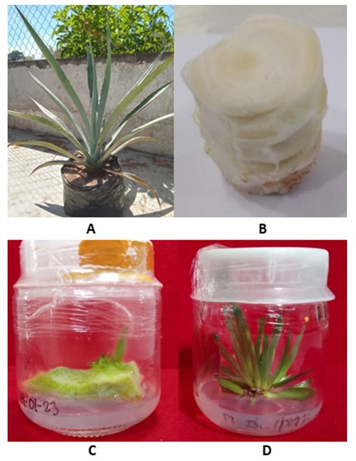
Figure 1 shows the condition of the plants in stage 0 in the nursery (Figure 1a), the stems that were taken from the nursery to the laboratory (Figure 1b), the stem tissues that were established in the culture medium in stage I (Figure 1c), and adventitious shoots that formed in stem tissues via organogenesis (Figure 1d).
When in vitro culture begins with tissues from the stem, which is a plant structure that in the nursery is in close contact with the soil and whose tissues harbor populations of endogenous microorganisms, the efficiency of treatments to eliminate microorganisms is limited. Ríos-Ramírez et al. (2018) reported that, when micropropagating A. angustifolia, in stage I, between 38.7 and 83.4% of the cultures were contaminated; for their part, López-Acevedo et al. (2018) report that 29.8% of the stem tissues they established in vitro were aseptic, viable, with adventitious shoot formation.
The works cited, as well as the present one, align with Debergh and Maene (1981) on the importance of stage 0 of prior management of plants in nurseries to reduce the incidence of contamination of plant tissues when they are established in vitro. Analyses of variance (Table 1) show that the propagation stages had highly significant differences in their effects (p ≤ 0.01) on the percentages of aseptic cultures obtained, contaminated cultures obtained and viable cultures obtained and significant effects (p ≤ 0.05) on the number of shoots formed in the explants.
| SV | DF | Arcsin aseptic (%) | Arcsin contaminated (%) | Arcsin viable (%) | NS |
|---|---|---|---|---|---|
| Treat | 3 | 9 980** | 10 633** | 10 679.91** | 0.77* |
| Error | 49 | 256.13 | 164.21 | 123.09 | 0.17 |
| Total | 52 |
As agave tissues age, it becomes difficult to cut them because their fibers have thicker cell walls than those in young stems. Likewise, the probability of having endophytic microorganisms in their tissues increases, so this factor was considered. During the first 10 days of incubation of stage I cultures, contaminating microorganisms of the genera Fusarium and Aspergillus, as well as gram-negative bacteria, were diagnosed in the tissues cultured in vitro; the evidence shows that, of the total agave stem tissues that were established in vitro, only 3.68% and 8.73% of the stem tissues in treatment 1 and treatment 2 were aseptic, respectively.
Nevertheless, some cultures of stem tissues that became contaminated remained viable and shoot organogenesis occurred, so these stem tissues were subjected to treatment 4 and microorganisms were eliminated in 52% of them (Table 2).
[i] T1= explants of two-year-old plants; T2= explants of five-year-old plants, for the establishment of aseptic cultures; T3= aseptic culture shoots; T4= shoots obtained in vitro, contaminated, which were superficially disinfected and established in culture medium with antibiotics; C= percentages of contaminated shoots; VAC= percentages of viable, aseptic cultures; NNS=number of new shoots. The data shows the average ±standard error. In each column, averages with the same letter are not significantly different (Tukey, 0.05).
Microbial contamination in stage I was due to endogenous microorganisms present in stem tissues, despite the application of fungicides and antibiotics in the nursery (stage 0). The surface disinfection used was ineffective against these internal contaminants, as the disinfectants act superficially. Contamination due to improper handling, poor sterilization of materials, or culture media is ruled out. Similarly, Sánchez-Cuevas and Salaverria (2004) point out that in strawberries, pubescence and contact with the soil favor the presence of endophytes that are difficult to eliminate when establishing in vitro cultures.
Aseptic, viable stem tissue cultures obtained in stage I that showed shoot formation were transferred to a new culture medium. After eight weeks, 89.88% of the cultures maintained their aseptic condition, 100% were viable and all formed an average of 1.07 new shoots (Table 2). The high percentage of viable cultures in which new shoots formed in stage II is because the plant materials are no longer subjected to the surface disinfection procedure, since this procedure, which was applied to the plant tissues in stage I to eliminate microorganisms, also damages the plant material.
When there was organogenesis of adventitious shoots from stem tissues in stage I and these cultures were contaminated with bacteria, they were extracted from the culture medium to undergo treatment 4, which included superficial disinfection, and then established in a culture medium to induce organogenesis, but the medium also contained the antibiotics chloramphenicol (100 mg L-1) and ampicillin (0.1 g L-1). After 20 days of incubation, 52.5% no longer showed fungal or bacterial growth, which is significantly lower (Tukey, 0.05) than the contamination of cultures in stage I. The antibiotic doses used in treatment 4 were determined in trials prior to this experiment. When chloramphenicol and ampicillin were added to the culture medium at concentrations higher than those indicated as effective, they produced phytotoxicity, causing tissue necrosis.
In in vitro cultures of Agave angustifolia, the level of microbial contamination varies with the propagation stage. In stage I, most contaminations (91.2-96.3%) are from endogenous microorganisms present in stem tissues, which are not eliminated by fungicides and antibiotics in the nursery or by surface disinfestation. However, in adventitious shoots already formed but contaminated with bacteria, an additional disinfection treatment plus establishment in a culture medium containing antibiotics allowed 52.5% of the explants to be recovered in an aseptic condition.
The origin of the plant material is key to the success of in vitro propagation. When using cultures that were aseptic in stage I and the clusters of shoots were subdivided for stage II (propagule multiplication), contamination was drastically reduced to only 10.1%, all cultures remained viable, and new shoots formed.
The data in the paper were obtained with the support of the Secretariat of Science, Humanities, Technology, and Innovation (SECIHTI), by its Spanish acronym, Mexico, which granted the first author a national scholarship for postgraduate studies.
Cruz-Hernández, H.; Enríquez-Valle, J. R.; Velasco-Velasco, V. A.; Ruiz-Luna, J.; Campos-Ángeles, G. V. y Aquino, D. E. 2013. Nutrimentos y carbohidratos en plantas de Agave angustifolia Haw. y Agave karwinskii Zucc. Revista Mexicana de Ciencias Agrícolas. 6(4):1161-1173. https://www.scielo.org.mx/pdf/remexca/v4nspe6/v4spe6a8.pdf.
Domínguez-Rosales, M. S.; González-Jiménez, M. L.; Rosales-Gómez, C.; Quiñones-Valles, C.; Delgadillo, D. S.; Mireles-Ordaz, S. J. y Pérez-Molphe, B. E. 2008. El cultivo in vitro como herramienta para el aprovechamiento, mejoramiento y conservación de especies del género agave. Investigación y Ciencia. Universidad Autónoma de Aguascalientes. 41:53-62. https://dialnet.unirioja.es/servlet/articulo?codigo=6104556.
Jarquín-Rosales, D.; Enríquez-Valle, J. R.; Alpuche-Osorno, J. J.; Rodríguez-Ortiz, G.; Martin, M. P. and Campos-Ángeles, G. V. 2022. The effects of fertirrigation and Azospirillum brasilense inoculation on photosynthetic compounds of Agave angustifolia. Australian Journal of Crop Science. 16:162-168. Doi: http://doi.org/10.21475/ajcs.22.16.01.p3280.
López-Acevedo, L.; Merino-Pérez, Y. E.; Enríquez-Valle J. R.; Rodríguez-Ortiz, G. and Lagunas-Sánchez, Z. C. 2018. Organogénesis in vitro en tejidos de tallo de Agave marmorata y Agave angustifolia. Revista Mexicana de Agroecosistemas. 5(2):20-27. https://revistaremaeitvo.mx/index.php/remae/article/view/165.
Miguel-Luna, M. E.; Enríquez-Valle, J. R.; Velasco-Velasco, V. A.; Villegas-Aparicio, Y.; Carrillo-Rodríguez, J. C. y Rodríguez-Ortíz, G. 2013. Composición del medio de cultivo y la incubación para enraizar brotes de Agave. Revista Mexicana de Ciencias Agrícolas. 4(6):1151-1159. https://www.scielo.org.mx/pdf/remexca/v4nspe6/v4spe6a7.pdf.
Obledo-Vázquez, E. N.; Flores-Verduzco, N. y Cervantes-Martínez, J. 2004. Detección del efecto de un extracto vegetal antimicrobiano sobre plantas de agave (Agave tequilana Weber var. azul) Cultivadas in vitro utilizando fluorescencia inducida por láser (LIF). Revista Mexicana de Fitopatología. 22(3):328-332. https://www.redalyc.org/pdf/612/61222302.pdf.
Pérez-Pazos, J.; Rosero, A.; Cardinale, M. and Gámez, R. 2023. Development of control strategies for bacteria and fungi associated with a micropropagated new cultivar of orange-fleshed sweet potato (Ipomoea batatas cv. Agrosavia-Aurora). Hortic. Environ. Biotechnol. 64:859-875. https://doi.org/10.1007/s13580-023-00521-2.